Get the latest news, inspiring stories, upcoming events, and valuable support services delivered straight to your inbox.
ALL NEWS
Filter by type

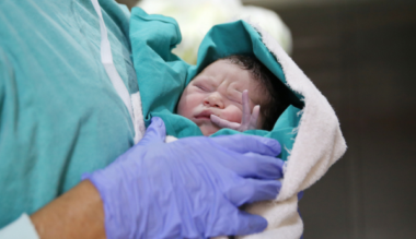
Every newborn baby in England to be screened for SMA
July 16, 2026
PIP ‘not fit for purpose’, Timms Review says
July 9, 2026

Collaborating with breast cancer awareness charity CoppaFeel!
June 25, 2026
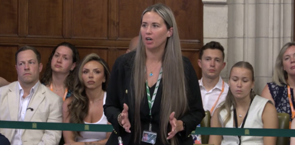
SMA newborn screening debated in Parliament
June 23, 2026
New grant fund available for home equipment
June 12, 2026
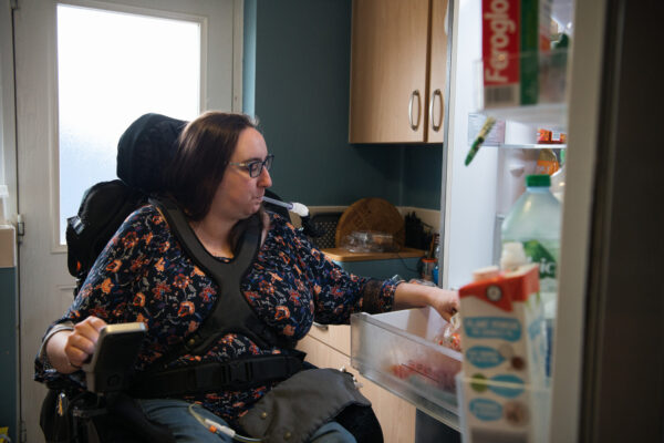
Translarna not accepted for routine use on the NHS in Scotland
June 8, 2026
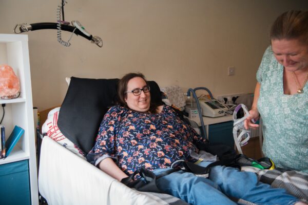
Sign our petition for a benefits system that supports everyone living with a muscle wasting condition
June 2, 2026
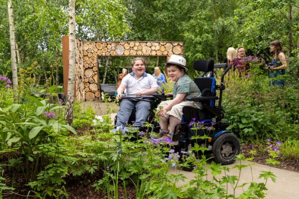
Our RHS Chelsea Flower Show garden officially opens at Glasgow’s Hospice
May 27, 2026
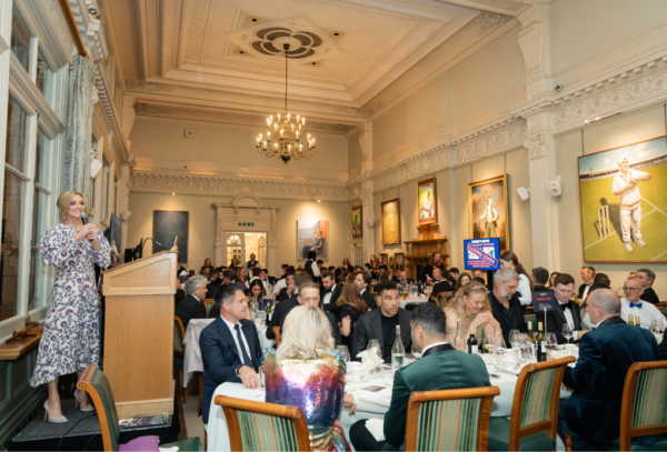
Celebrity Sports Quiz 2026 raises over £97,000
May 27, 2026
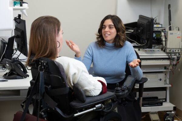
A decade of progress: from zero to ten treatments for muscle wasting conditions
May 27, 2026
Opening applications for our 2026 Improving Quality of Life research grant scheme
May 26, 2026
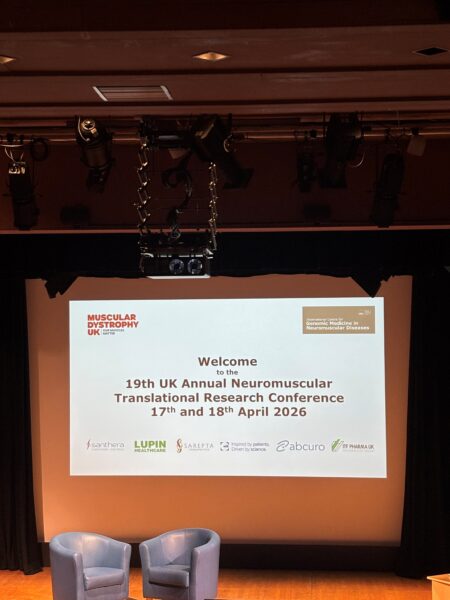
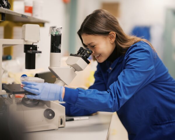
The latest research into muscle wasting conditions at the 19th UK Neuromuscular Translational Research Conference
May 22, 2026