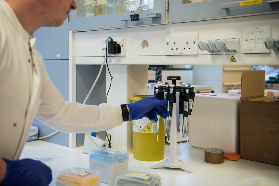
Professor Jordi Diaz-Manera and colleagues at Newcastle University and institutes in Barcelona and Madrid, carried out a study to see if magnetic resonance imaging (MRI) can be used to tell dysferlinopathies and other muscle wasting conditions apart.
Using MRI to tell dysferlinopathy apart from other genetic muscle wasting conditions
19 July 2024
Grace Danielle Ifura writes…
Dysferlinopathies are a group of conditions caused by changes in the DYSF gene – the gene that produces dysferlin protein which is important in muscle repair following injury. These changes in the DYSL gene lead to unsuccessful repair of the muscle fibre, causing a loss of muscle fibre with a build-up of fibrotic (scar) and fat tissue in its place.
Challenges in diagnosis
Some dysferlinopathies are also part of the group of limb girdle muscular dystrophies (LGMD), such as LGMDR2 (formerly known as LGMD2B). LGMDs are a group of muscular dystrophies that affect proximal muscles, closest to the centre of the body, of the limbs and spread to other muscles as the condition progresses. However, people with different LGMDs often have similar characteristics, known as phenotypes, especially when the condition has progressed. So, the use of genetic studies, biopsies and imaging data could make it easier to differentiate between these different types of LGMDs.
Setting MRI-based criteria for dysferlinopathies diagnosis
A natural history study called the International Clinical Outcome Study of Dysferlinopathy (COS) was carried out over six years ago, which collected genetic, clinical, muscle function and radiological data from 182 patients diagnosed with a type of dysferlinopathy. Magnetic resonance imaging (MRI) was used to identify and measure the pattern of fat replacement in groups of muscles completely or partially replaced by fat due to muscle damage, and muscles less affected or not affected at all. The results from this study were used to create a set of criteria for diagnosis.
A new study to differentiate between muscle wasting conditions during diagnosis
Given the need for easier and quicker diagnosis of specific conditions, in 2022 we awarded a grant to Professor Jordi Diaz-Manera to study the use of MRI-based criteria to differentiate between different genetic muscle wasting conditions.
The 182 patients from the COS study, mentioned above, were participants along with an additional 1000 people living with 10 other limb girdle muscle weakness conditions. These participants were collected through a network of professionals called the MYO-Guide Consortium. MYO-Guide is a computer modelling tool that can suggest a diagnosis of 10 conditions with high accuracy based on the muscle MRI analysis.
The amount of fat in the pelvis, thigh and lower leg was measured, and the level of fat replacement per region was analysed using the criteria for diagnosis set in the COS study.
Study results
Researchers found that patterns of muscle fatty replacement in the legs of people with dysferlinopathy are similar to patterns described for other muscle wasting conditions, such as FKRP-related myopathy, anoctamin-5 myopathy and calpainopathy. This result was expected, as they are all clinically similar. However, MRI results could show that oculopharyngeal muscular dystrophy (OPMD) and Duchenne muscular dystrophy (DMD) are different to dysferlinopathy in terms of muscle fatty replacement.
What do these mean?
The researchers created a set of rules, called an algorithm, to see if they could tell dysferlinopathy apart from other conditions using MRI. The rules were really sensitive, which means that they could detect most cases and didn’t miss many cases. However, the rules were not terribly specific because several muscle wasting conditions show similar patterns of muscle fat replacement. Therefore, a diagnosis of dysferlinopathy can’t be made using MRI alone. But the researchers say that this method could be used to rule out other conditions that show different muscle fat replacement patterns, such as oculopharyngeal muscular dystrophy (OPMD) or Duchenne muscular dystrophy (DMD).
For now, it remains important to use MRI in combination with other clinical approaches, such as observing clinical features of people living with the conditions and genetic testing. And a more complicated set of rules is needed to differentiate between muscle wasting conditions to help diagnosis.
STORIES LIKE THIS

Update on givinostat – and what it means for families
March 16, 2026

SMA explained: Reflecting on Jesy Nelson’s news
January 5, 2026
Discover our bold new ten-year strategy
September 17, 2025