As part of the pathway to diagnosis, your doctor may request a muscle biopsy to help with diagnosis. This is usually after a physical examination, blood tests, and possibly an electromyography (EMG).
Understanding muscle biopsies
Skip to section
What is a muscle biopsy?
A muscle biopsy is a procedure in which a small sample of muscle – usually about the size of an orange pip – is taken and examined under a microscope. It is a minor procedure and is usually done in outpatients under local anaesthetic or as a day case under local or general anaesthetic.
What does a muscle biopsy involve?
A muscle biopsy can be taken from almost any muscle, but a doctor will take the sample from muscle that is affected by your suspected condition – but is not severely wasted.
A muscle biopsy does not increase any muscle weakness and the muscle will regrow in time.
Types of muscle biopsy
There are two types of muscle biopsy: a needle biopsy and an open biopsy. Both types of biopsies have advantages and disadvantages, and different hospitals often prefer to use different methods.
1. Needle biopsy
A needle biopsy is a specialised procedure and is only performed in outpatients or as a day case at a few centres in the UK. It involves inserting a needle, about 5mm in diameter, into your muscle. When the needle is removed, it has inside it a small sample of muscle that is taken for analysis. The needle biopsy scar is very small and is closed with sterile strips and a plaster. You won’t need stitches.
2. Open biopsy
An open biopsy involves making a cut in your skin to remove a muscle sample. The cut is usually just a few centimetres long and once the sample is taken, the cut is closed with stitches.
The scar from an open biopsy is bigger than from a needle biopsy, because a bigger muscle sample has been removed. Sometimes a bigger sample is necessary and makes it less likely you will need a second biopsy.
Are there any risks with muscle biopsies?
The risks associated with muscle biopsies are very small. There is a minor risk of muscle damage and infection, and you’ll often have a patch of numbness around the scar, which may last for a few weeks.
Is there an alternative to a muscle biopsy?
A muscle biopsy is a standard procedure when doctors are investigating whether you might have a muscle wasting condition.
Molecular genetic testing is available for some conditions, so in those cases a muscle biopsy may not be necessary. Doctors usually rule out any of these conditions first by analysing a blood sample. They will then decide whether to do a muscle biopsy too.
What happens after the muscle biopsy?
Your muscle sample is sent to the laboratory, where most of it is frozen. Very thin slices are cut and stained with various dyes and examined under a microscope.
A small piece of your muscle sample may be put in a preservative so it can be examined at extremely high magnification using an electron microscope (see Electron microscopy below).
Some of the muscle sample may also be used for other types of studies, which also help to explain what is happening inside your muscle.
If you agree, any parts of the sample which have not been used can be stored so they can be studied for research purposes. The pathologist who analyses your muscle sample will examine it in different ways depending on which muscle wasting condition your doctor suspects you may have.
Different techniques of examining a muscle sample
There are different techniques used to examine a muscle sample. These are explained below.
Histology
This involves looking at the overall appearance and structure of your muscle cells so the pathologist can look for characteristics that are specific to certain conditions. The muscle is dyed using various chemicals so that the different structures show up under the microscope.
Histochemistry
This technique also uses chemical dyes but looks at the activity of chemicals within your muscle fibres. This is important when diagnosing metabolic disorders. Histochemistry also reveals the characteristics of your muscle fibres. Certain changes can help to identify particular conditions.
Immunohistochemistry
This procedure uses antibodies that bind to a specific protein and can show the presence or absence of important proteins within the muscle.
When antibodies are tagged with a marker, they can be seen under the microscope and can show if a protein is in the wrong place or is absent or reduced in amount.
This is important for several muscular dystrophies, such as Duchenne muscular dystrophy, which is caused by the absence of the dystrophin protein.
Electron microscopy
Electron microscopy allows high magnification of each muscle cell, making it easier to see structural abnormalities. This is relevant for conditions such as nemaline myopathy, where the diagnosis is based on the presence of rod structures in the muscle. Some abnormal features are visible only with the high magnification provided by electron microscopy.
What are pathologists looking for?
There are a number of things a pathologist will be looking for when they examine your muscle sample, to help them come to a diagnosis.
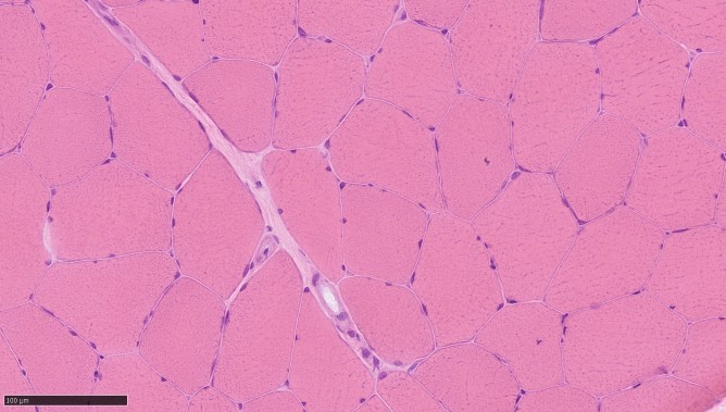
Appearance of fibres
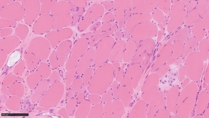
Healthy muscle has a characteristic appearance, and is made up of closely packed fibres, which are more or less evenly sized. See Figure 1 below.
Muscle affected by a muscle wasting condition looks different from normal muscle, which you can see in Figure 2. These differences vary between conditions.
There are two types of muscle fibre: type 1 and type 2.
In some conditions, the muscle fibres are smaller or larger than they should be. They may be damaged, or the proportion of type 1 to type 2 fibres may be unbalanced. In some cases, only one type of fibre may be affected.
Important proteins
Muscle fibres are built from a number of different essential proteins. If some of these proteins are missing, in the wrong place, or there are too many or too few of them, this may cause problems with the muscle.
Buildup of substances in your muscle
There are many different chemical pathways within muscle tissue that can be affected in muscle wasting conditions. These result in changes in the amounts of key substances in your muscle.
One example of this is glycogen, which is an important energy storage molecule. In healthy muscle, glycogen is broken down by a pathway involving several proteins called enzymes.
Enzymes control the speed of chemical reactions in your body, but in some muscle wasting conditions, one of these enzymes is missing or abnormal.
This results in a build-up of glycogen in your muscle, which can be seen under the microscope with certain histochemical dyes. Special biochemical studies may be needed to identify the exact problem. A build-up of certain proteins in muscle fibres can occur in some conditions, and these can be identified by certain antibodies.
Structural changes
Some conditions are diagnosed by the presence of structural abnormalities within the muscle.
For example, muscle affected by core myopathy (including central core disease and multiminicore disease) has characteristic core structures.
In mitochondrial myopathies, structures called mitochondria, which convert food into energy, contain faulty proteins that disrupt their function. These can sometimes be seen under a microscope or revealed with special biochemical studies.
Distribution
It is important to look at the distribution of any abnormalities within the muscle. This can sometimes, but not always, give an indication of the severity, and the rate of progression of a condition.
How long do I have to wait for the results?
How long you will have to wait for your results will depend on the number of tests you need and the date of your next appointment with your consultant.
When your consultant has received the results of your muscle biopsy, they will invite you to an appointment to discuss the results and the follow-up. What happens next depends on the results of your tests and your consultant will discuss this with you.
We’re here to support you
Our support services
Webinars, Information Days, and support groups for our muscle wasting community. Our life-changing support is here for you.
Call our helpline
Information
Advice for living with or caring for someone with a muscle wasting condition.